Diagram of human eye anatomy with label 1848847 Vector Art at Vecteezy
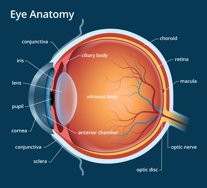
1. Conjunctiva The conjunctiva is the membrane covering the sclera (white portion of your eye). The conjunctiva also covers the interior of your eyelids. Conjunctivitis, often known as pink eye, occurs when this thin membrane becomes inflamed or swollen. Other eye disorders that affect the conjunctiva include:

Brain Post How Big is Your Blind Spot? Human eye diagram, Eyeball structure
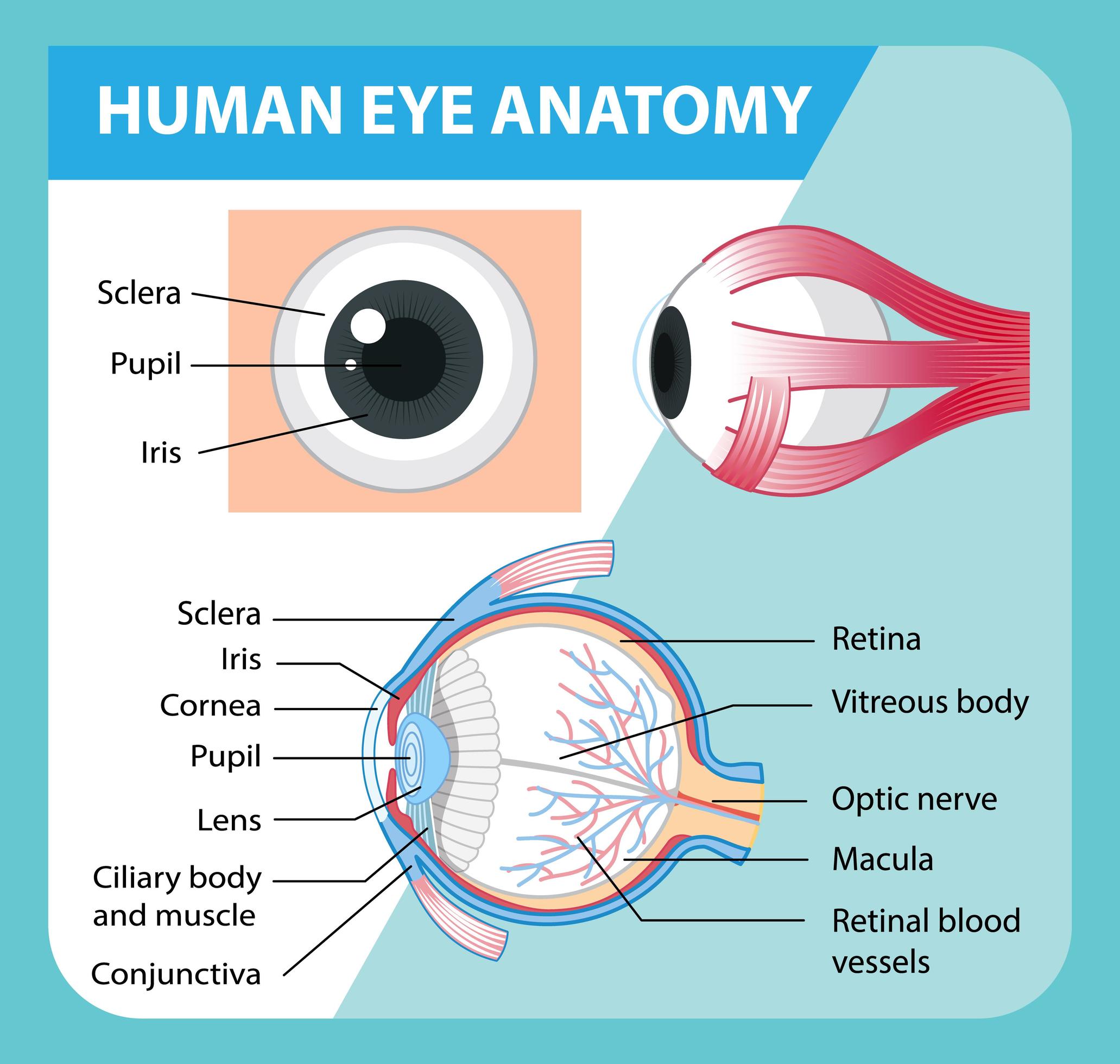
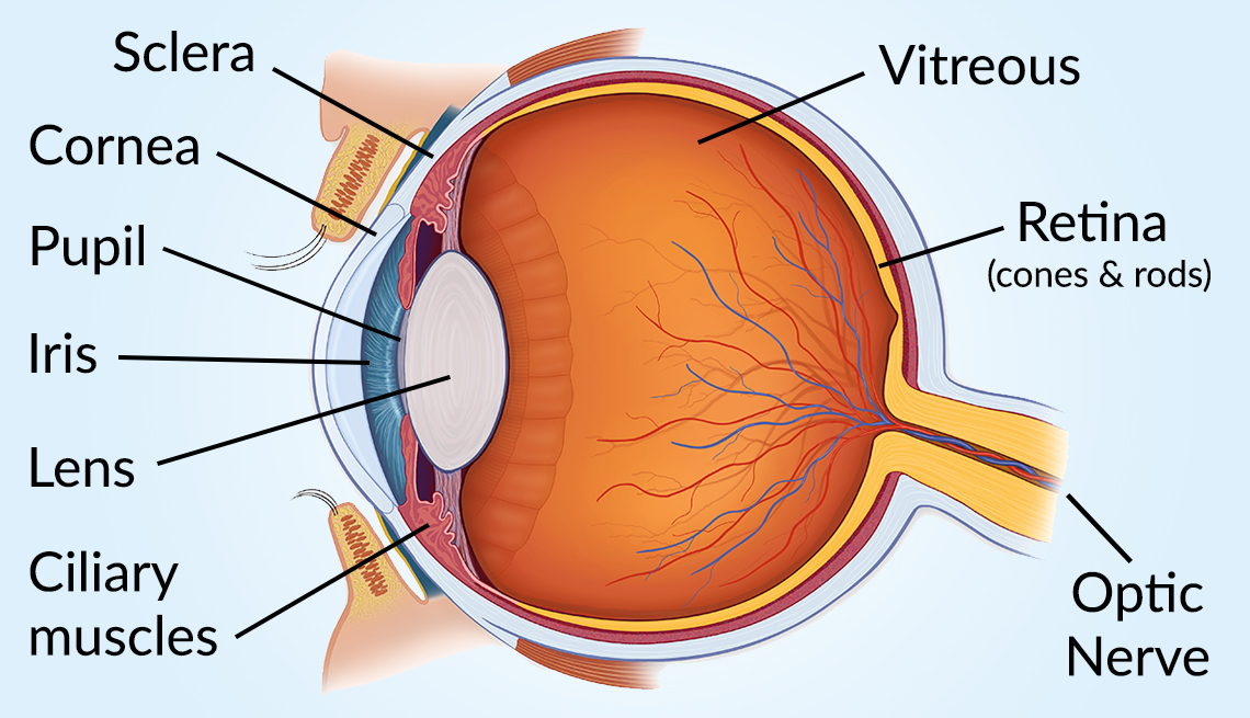
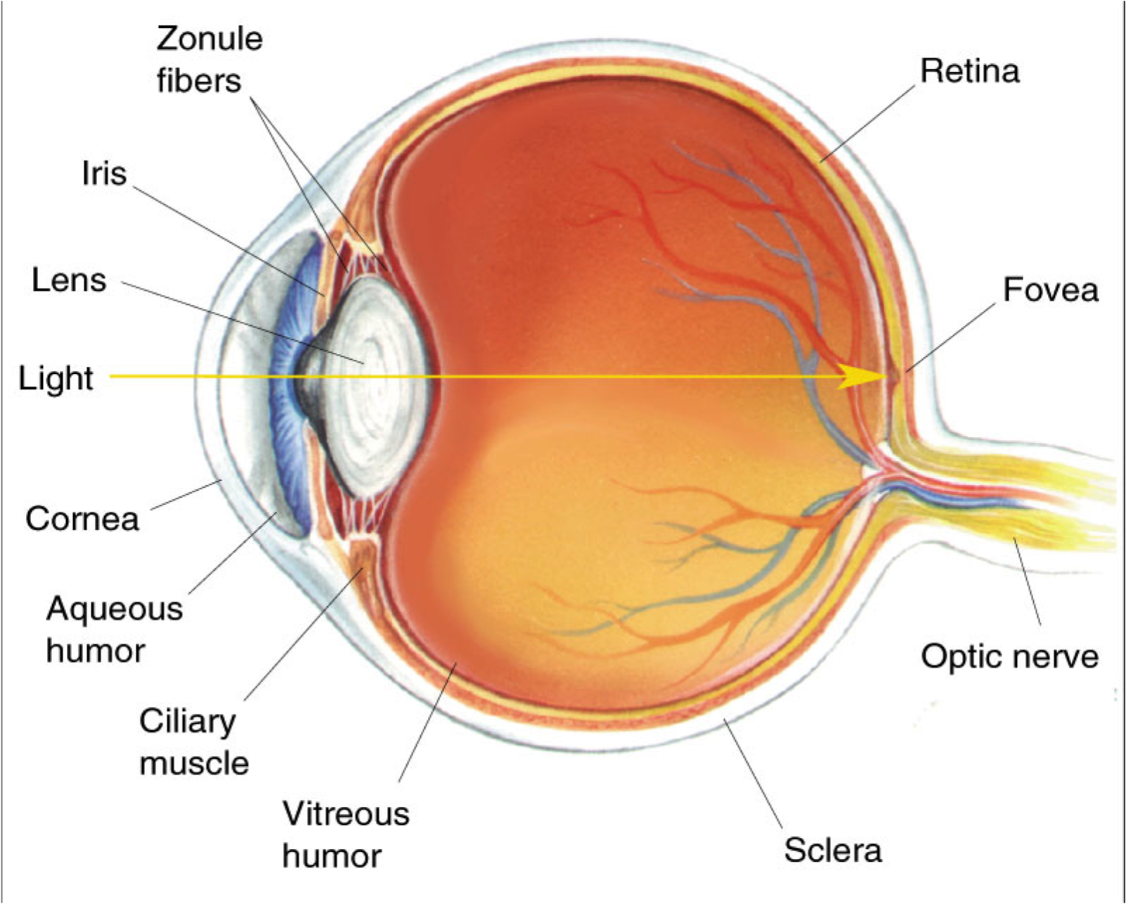
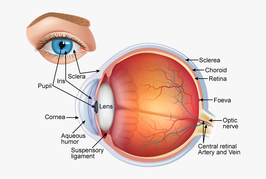
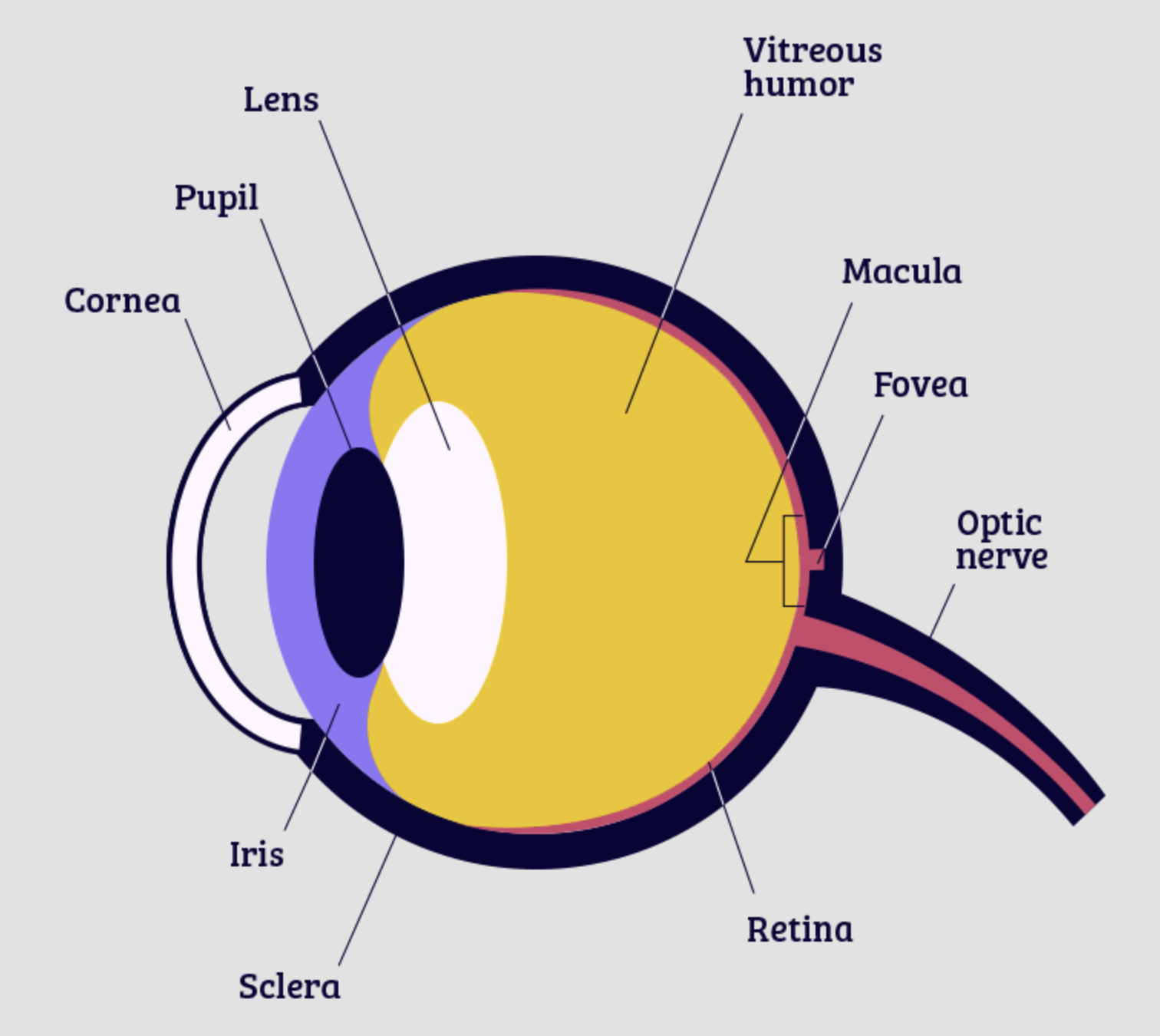
Light is focused primarily by the cornea - the clear front surface of the eye, which acts like a camera lens. The iris (colored part) of the eye functions like the diaphragm of a camera, controlling the amount of light reaching the retina by automatically adjusting the size of the pupil (aperture). The eye's crystalline lens is located.
Vision and Eye Diagram How We See
Cornea: The clear, dome-shaped tissue covering the front of the eye. Fovea: A tiny pit located in the macula of the retina that provides the clearest vision of all. Iris: The colored part of the eye that controls the amount of light that enters the eye by changing the size of the pupil. Lens: A crystalline structure located just behind the iris.
Eye Diagram Cliparts.co
Diabetes Healthy ANATOMY and Eyes OF THE AND ITS FUNCTION Toolkit Parts of the Eye Vision is wonderful, but you could lose To understand it if you eye have problems, diabetes. it is helpful to know the different parts of the eye. Please refer to the back of this handout for descriptions of their functions. The main parts of the eye— Optic 3

Human Eye Anatomy Parts of the Eye and Structure of the Human Eye
Download. English: Parts of the Eye (PDF 603.5 KB) Spanish: Las partes del ojo (PDF 897.7 KB) Check out this fact sheet to see a labeled diagram of the eye and learn about the different parts of the eye.

eye diagram Discovery Eye Foundation
6 min read Your eye is a slightly asymmetrical globe, about an inch in diameter. The front part (what you see in the mirror) includes: Iris: the colored part Cornea: a clear dome over the iris.
HUMAN EYE (STRUCTURE, IMAGE FORMATION AND DIFFERENCE BETWEEN RODS AND CONES) « SimpleBiology
Eyelid anatomy Lacrimal gland Eye muscles Eyeball Outer layer Middle layer Inner layer Blood supply of the eye Nerves of the eye Sources + Show all Bones of the orbit The bony orbit is made out of seven bones, which include the maxilla, zygomatic bone, frontal bone, ethmoid bone, lacrimal bone, sphenoid bone and palatine bone.
Human Eye Labelled Diagram , Free Transparent Clipart ClipartKey
Eye Diagram Handout Parts of the Eye To understand eye problems, it helps to know the different parts that make up the eye and the functions of these parts. Here are descriptions of some of the main parts of the eye: Cornea: The cornea is the clear outer part of the eye's focusing system located at the front of the eye.
/GettyImages-695204442-b9320f82932c49bcac765167b95f4af6.jpg)
Structure and Function of the Human Eye
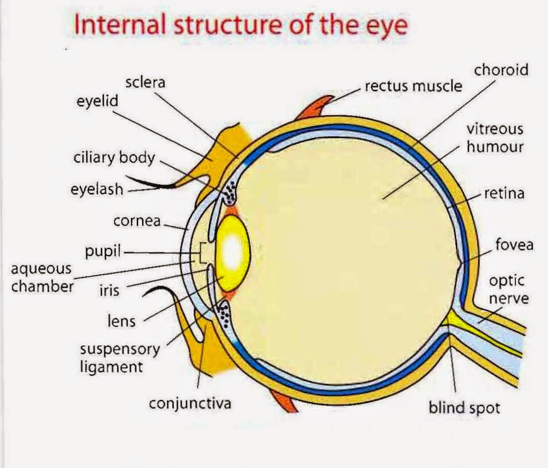
A small, red portion of the corner of the eye that contains modified sebaceous and sweat glands. Choroid. The thin, blood-rich membrane that lies between the retina and the sclera and is responsible for supplying blood to the outer portion of the retina. Ciliary body. The part of the eye that produces aqueous humor. Cornea.
About the Eye National Eye Institute
The structures and functions of the eyes are complex. Each eye constantly adjusts the amount of light it lets in, focuses on objects near and far, and produces continuous images that are instantly transmitted to the brain. The orbit is the bony cavity that contains the eyeball, muscles, nerves, and blood vessels, as well as the structures that.

File1413 Structure of the Eye.jpg Wikimedia Commons
Diagram Of Eye The human eye is responsible for the most important function of the human body, the sense of sight. It consists of several distinct parts that work in coordination with each other. The most common eye diseases include myopia, hypermetropia, glaucoma and cataract.

Human Eye Anatomy, Structure and Function
Labelling the eye — Science Learning Hub Interactive Labelling the eye Interactive Add to collection Use this interactive to label different parts of the human eye. Drag and drop the text labels onto the boxes next to the diagram. Selecting or hovering over a box will highlight each area in the diagram. Cornea Lens Retina Optic nerve Pupil Schlera
Human Eye Anatomy, parts and structure Online Biology Notes
Muscles in the iris dilate (widen) or constrict (narrow) the pupil to control the amount of light reaching the back of the eye. Directly behind the pupil sits the lens. The lens focuses light toward the back of the eye. The lens changes shape to help the eye focus on objects up close.
:max_bytes(150000):strip_icc()/eye-conjunctiva-871453538-5a26c6ad22fa3a0037d5edad.jpg)
How the Human Eye Works (Structure and Function)
The main parts of the human eye are the cornea, iris, pupil, aqueous humor, lens, vitreous humor, retina, and optic nerve. Light enters the eye by passing through the transparent cornea and aqueous humor. The iris controls the size of the pupil, which is the opening that allows light to enter the lens. Light is focused by the lens and goes.

Human eye Extraocular Muscles Britannica
Human Eye Diagram: Contrary to popular belief, the eyes are not perfectly spherical; instead, it is made up of two separate segments fused together. Explore: Facts About The Eye To understand more in detail about our eye and how our eye functions, we need to look into the structure of the human eye. Recommended Video: 1,221

Diagram showing the different parts of the eye Parts of the eye, Eye health, Free homeschool
The eye is protected from mechanical injury by being enclosed in a socket, or orbit, which is made up of portions of several of the bones of the skull to form a four-sided pyramid, the apex of which points back into the head.
